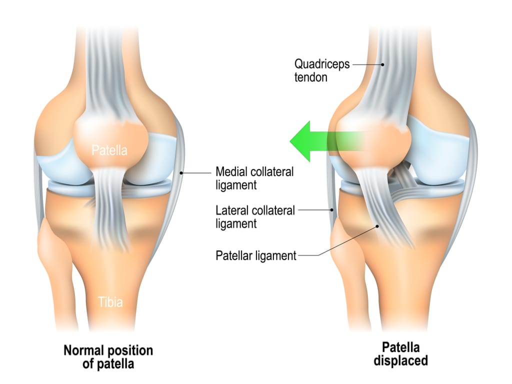
A Patellar dislocation is characterized by displacement of the patella (knee cap) and tearing of the connective tissue surrounding the patella.
The patella is located at the front of the knee and lies within a groove at the front of the femur (thigh bone). The tendon of the quadriceps muscle (the muscle at the front of the thigh) wraps the patella and attaches to the top end of the tibia (shin bone). Connective tissue called the patella retinaculum attaches the knee cap in place upon the femur. Occasionally the patella may be forced out of its normal position, known as a patellar dislocation.

What are the symptoms of a patellar dislocation?
Typically, a patellar dislocation presents as a sudden, intense pain at the front of the knee during injury. Pain is usually associated with the feeling that the knee is going to ‘give way’ or the feeling of something ‘popping out’. There may be a visible deformity of the knee compared to the unaffected side. There may be a rapid onset of knee swelling within the first 1-2 hours following injury. Once the patella has returned to its original position following dislocation, individuals typically experience an ache that may increase to a sharper pain with movement. Pain is generally experienced during activities that bend or straighten the knee particularly whilst weight bearing. For example, going up and down stairs or hills, squatting, lunging, running or jumping. The borders of the patella may be tender to touch. Patients may also notice a clicking or grinding sound when bending or straightening the knee.
How can physiotherapy help?

